Menu
Menu


Context & Study Overview
Metastatic castration-resistant prostate cancer (mCRPC) remains an incurable disease, despite recent therapeutic advances. [177Lu]Lu-PSMA radioligand therapy (RLT) has become an important treatment option, providing meaningful clinical benefits in selected patients. However, approximately 30% fail to respond to this therapy, highlighting the need for reliable pre-therapeutic predictive biomarkers to better assess individual prognosis.
Although several clinical and imaging prognostic factors have been identified, accurately estimating patient outcomes remains challenging. In this context, machine learning (ML) models offer a promising approach by integrating multiple data modalities to enhance prognostic assessment.
This retrospective study aimed to develop and evaluate ML models capable of predicting overall survival (OS) in patients with mCRPC treated with [177Lu]Lu-PSMA RLT following progression on androgen receptor pathway inhibitors (ARPIs) and taxane therapy.
By combining baseline clinical, biochemical, genomic, and imaging parameters, including PSMA-PET radiomic features, the researchers investigated the potential of a multimodal AI (MMAI) approach to improve patient risk stratification in a real-world setting.
Dataset and Methods
This monocentric retrospective study included mCRPC patients treated with [177Lu]Lu-PSMA RLT at Institut Bergonié, in Bordeaux, France, between January 2022 and March 2024. Of the 150 patients initially screened, 102 met the study criteria and were included in the final analysis.
A total of 80 pre-therapeutic clinical, biochemical, genomic, and radiological parameters were collected from electronic health records (EHR). Additionally, approximately 280 radiomic features were extracted from baseline [68Ga]Ga-PSMA-11 PET/CT scans, following the Image Biomarker Standardization Initiative (IBSI) guidelines.
Out of a total of approximately 340 collected features, 22 were selected for the final analysis, encompassing:
Three families of survival ML models were trained and evaluated: Cox proportional hazards models with LASSO or ridge regularization, random survival forests, and gradient-boosted trees.

Figure 1. Patient selection and feature processing workflow for ML model development.
Three predictive models were subsequently developed and evaluated using the same nested cross-validation framework:
The primary outcome assessed was OS, defined as the time from initiation of [177Lu]Lu-PSMA RLT to death from any cause.
Key Findings
The median OS across the 102 patients included in the study was 11.2 months (95% CI 7.4-12.8).
The best-performing ML model was a Cox proportional hazards model with LASSO regularization. Among the three evaluated models, the full multimodal model, integrating all available data modalities, achieved the highest 12-month AUC (0.778, 95% CI 0.649-0.908).
The imaging-only model also demonstrated strong performance, achieving an AUC comparable to that of the clinical and biological model (0.715 and 0.766, respectively; Table 1). This highlights the substantial prognostic value of PSMA-PET imaging, including radiomic features, even without extensive clinical and molecular information.
All three models demonstrated strong calibration and successfully stratified patients into high- and low-risk subpopulations with significantly different prognoses.
Table 1. Performance of the three models on selected features, assessed by 12-month AUC, and their ability to stratify patients into subpopulation risk groups.
| Model | Selected features | 12-month AUC (95% CI) | HR, low- vs high-risk groups (95% CI) |
|---|---|---|---|
| Full Multimodal | All modalities | 0.778 (0.649-0.908) | 0.40 (0.26-0.62) |
| Clinical/Biochemical | Clinical and biochemical | 0.766 (0.644-0.888) | 0.42 (0.27-0.65) |
| Imaging/Radiomic | PET | 0.715 (0.594-0.836) | 0.46 (0.29-0.71) |
Legend: AUC, area under the curve; HR, hazard ratio
Prognostic factors associated with OS were also identified. Notably, higher intralesional homogeneity emerged as an independent prognostic factor associated with shorter OS (HR 1.64, 95% CI 1.07-2.53; p = 0.025).
Furthermore, the multimodal model consistently outperformed individual established prognostic variables, including the presence of liver metastases and baseline SUVmean, demonstrating the added value of integrating multiple data modalities for more comprehensive prognostic assessment.
Conclusion and Clinical Implications
This study demonstrates the potential of AI-powered multimodal approaches to improve OS prediction in patients with mCRPC treated with [177Lu]Lu-PSMA RLT. By combining clinical, biochemical, genomic, and imaging features, the researchers developed a model capable of stratifying patients according to their expected survival outcomes.
Importantly, the findings highlight that PSMA-PET imaging alone can provide robust prognostic information, performing comparably to models based on clinical and biological variables. By extracting quantitative information beyond conventional visual image assessment, radiomic analysis can reveal additional tumor characteristics and heterogeneity patterns associated with patient outcomes.
These findings are particularly relevant in heavily pretreated patients, where therapeutic options are limited and a better understanding of individual prognosis could help guide treatment management.
This study provides encouraging evidence for the role of multimodal AI in mCRPC care, while also highlighting important next steps towards clinical implementation. Given its retrospective, single-center design, prospective multicenter studies and external validation will be important to confirm the models’ generalizability. In the absence of a comparator treatment arm, the identified features should currently be considered prognostic rather than predictive of treatment benefit. Further validation and technical standardization could help pave the way for their integration into clinical decision-making.
As the PSMA RLT landscape continues to evolve towards earlier disease stages, the findings underscore the importance of fully exploiting PET-derived data and the potential of AI-powered multimodal approaches to unlock additional prognostic insights. Ultimately, these approaches could support improved patient stratification and more informed, personalized treatment strategies.
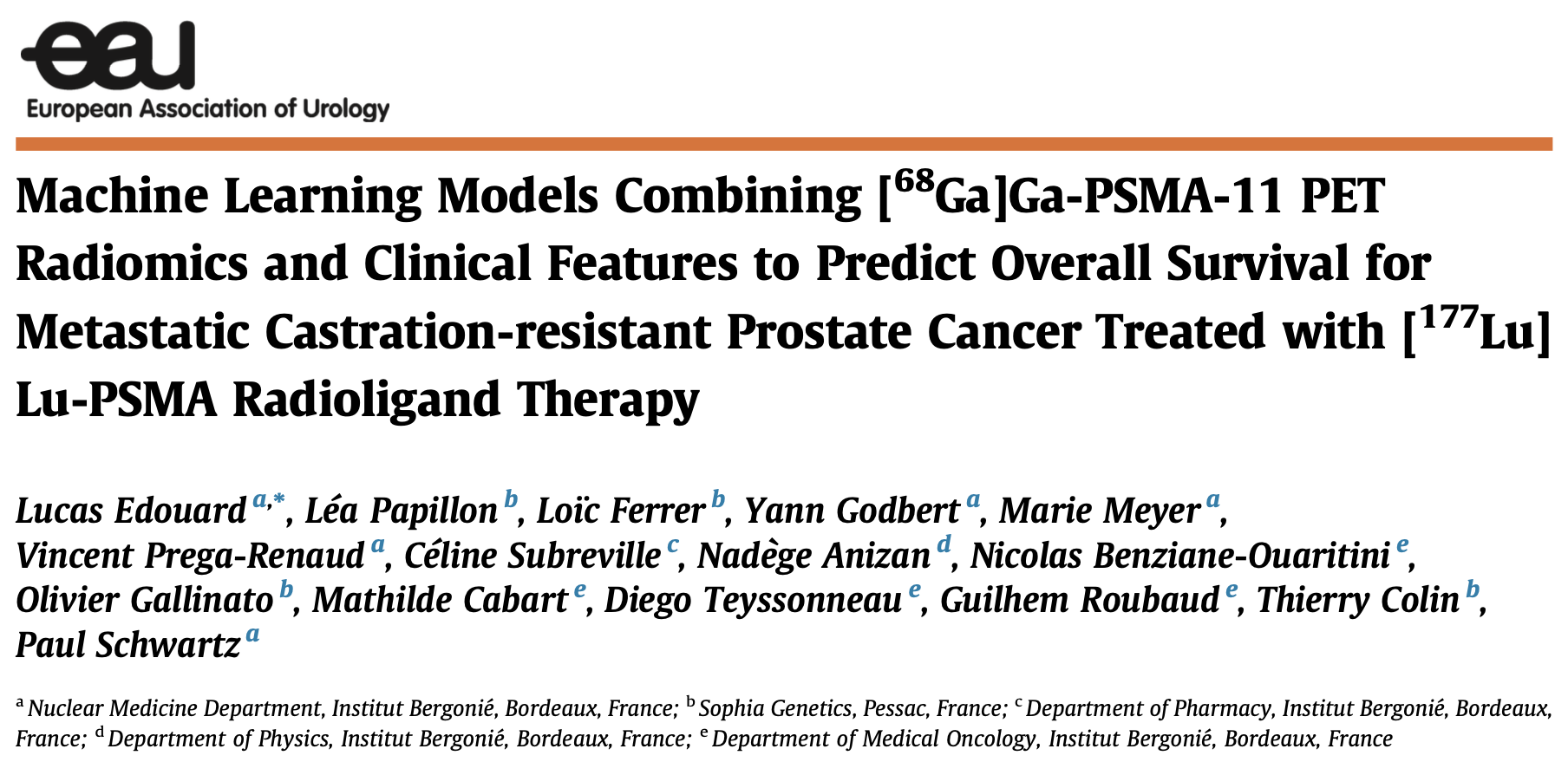
Edouard L, et al. Eur Urol Oncol. 2026. doi: 10.1016/j.euo.2026.09.015
Disclaimer
This project was executed by SOPHiA GENETICS in collaboration with Institut Bergonié, Bordeaux, France.
The study team leveraged the SOPHiA DDM™ for Multimodal Factory, including SOPHiA DDM™ for Radiomics and its proprietary algorithms, to perform this retrospective data analysis. If you want to know more about SOPHiA DDM™ for Multimodal, please visit our dedicated webpage.
SOPHiA DDM™ for Multimodal is for Research Use Only. May not be available for sale
SOPHiA GENETICS products are for Research Use Only and not for use in diagnostic procedures unless specified otherwise.
SOPHiA DDM™ Dx Hereditary Cancer Solution, SOPHiA DDM™ Dx RNAtarget Oncology Solution and SOPHiA DDM™ Dx Homologous Recombination Deficiency Solution are available as CE-IVD products for In Vitro Diagnostic Use in the European Economic Area (EEA), the United Kingdom and Switzerland. SOPHiA DDM™ Dx Myeloid Solution and SOPHiA DDM™ Dx Solid Tumor Solution are available as CE-IVD products for In Vitro Diagnostic Use in the EEA, the United Kingdom, Switzerland, and Israel. Information about products that may or may not be available in different countries and if applicable, may or may not have received approval or market clearance by a governmental regulatory body for different indications for use. Please contact us to obtain the appropriate product information for your country of residence.
All third-party trademarks listed by SOPHiA GENETICS remain the property of their respective owners. Unless specifically identified as such, SOPHiA GENETICS’ use of third-party trademarks does not indicate any relationship, sponsorship, or endorsement between SOPHiA GENETICS and the owners of these trademarks. Any references by SOPHiA GENETICS to third-party trademarks is to identify the corresponding third-party goods and/or services and shall be considered nominative fair use under the trademark law.